Posts

Showing posts from August, 2021

Macular Degeneraton | Symptoms | Causes | Diagnosis | Treatment

Image
Overview Macular degeneration is a disease that affects the retina. The retina is the layer at the back of the eye which helps us to see the world around us. Macular degeneration causes changes in central vision. Images that were once clear may appear blurred, Later dark spots may appear and enlarged, straight lines may become distorted or curved, colors may appear less vivid or darker. The eyes work together when one eye loses vision it may not be noticed because the other eye is still able to see each eye must be tested on its own to identify changes in vision. Symptoms The symptoms of macular degeneration may be found in other eye conditions. It is very important for an ophthalmologist to perform an examination to ensure that no, other conditions are present.  Dry macular degeneration affects the health of the retina. The retina is the lining of the eye that responds to light. Here a cross-section of the retina can be seen in the dry form of macular degeneration. Metabolic end p...

Colour blindness red, green |Test for colour blindness

Image
 Colour Blindness The inability of seeing color in a normal way is known as Colour blindness. Colour blindness is also known as color deficiency. It happens when someone did not recognize certain color. This usually happens between the color like red, green, and blue.  This diagnostic test will help to determine if you suffer from red-green color blindness. Can you identify the following numbers?  if you had difficulties in identifying the numbers you may be suffering from red-green color blindness and should consult with an optometrist. In the retina, we have two types of cells that detect light and color. They are rods and cones. Rods only detect light and dark which are sensitive to low light levels. whereas cones detect color and are concentrated near the center of vision. There are three types of cones that detect color: Red, Green, and Blue. Symptoms The symptoms of color blindness range from mild to severe. Most of the people did not recognize the color deficiency ...

6 Most common eye problems

Image
Eyes Problems Visibility of the world is under the eye so, it is the important sense organ of our body. In this 21st century, people are using mobile phones, computers, television for a long period of time. People are eating junk food, oily food. They are bringing new diseases day by day. Now talking about our eyes let's talk about  The most common problem of the eye.  Refractive error Conjunctivitis Cataract Glaucoma Diabetic Retinopathy Dry Eye Refractive error  Refractive error is the most common problem for many people. There are three types of refractive error include Hypermetropia: It is long-sightedness  Myopia: It is short-sightedness Astigmatism The inability to focus the image on the retina is due to blurred vision. It is also a serious problem for the eye if it is not treated on time. This disease can also lead to blindness. It can be treated by eyeglasses, contact lenses, or in some cases Surgery.  Conjunctivitis Any inflammation of the conjunctiva i...

Common Eye Problem in Children

Image
   The most common eye problem in children is Refractive error, Chalazion. Refractive Error There are three types of refractive error : Myopia Hypermetropia Astigmatism It causes blurred vision due to an inability to focus the image on the retina. If it is not corrected in time then it may be harmful to your child. As we all know our vision will be developed from childhood.  Refractive error:   The cornea and lens of the eye helps to focus. Refractive error is the vision problem that happens when the shape of the eye didn't keeps you focusing well.   Hypermetropia(+) Hypermetropia or long-sightedness is the refractive state of the eye wherein parallel rays of light coming from infinity are focused behind the retina with accommodation being at rest. Myopia(-) Myopia or short-sightedness is the refractive state of the eye wherein parallel rays of light coming from infinity are focused in front of the retina with accommodation being at rest. Astigmatism ...

Best Lens for Cataract Surgery

Image
  If you are ready for cataract surgery then at first you must know in this surgery your doctor will place a new artificial lens inside your eye. For sure you have to know about the lens,  which lens will be best for you. After all, you have to choose the best lens for the rest of your life. As we are talking about the lens,  There are 3  types of lens available  Monofocal lens Multifocal lens Trifocal lens What is lens? The lens is a transparent medium that has at least one curved surface. The lens is the most important part of the eyes.  During cataract surgery, your doctor will place a clear artificial lens inside your eye to sharpen light particles as they move through your eye. Most of the lens is made of silicone or acrylic. These lenses are also coated with materials that protect us from damage caused by the sun's rays. As you must discuss with your eye surgeon which lens will be best for your eyes and your vision. You have several choices ...

Lazy eye (Amblyopia) | Causes| Symptoms| Diagnosis| Treatment

Image
Lazy Eye (Amblyopia) A mblyopia occurs when there is a lack of vision in one eye because the eye and the brain are not working together. Leo Pia is an early childhood condition that develops because of abnormal visual experience that changes the nerve pathways between the retina and the brain when an individual has amblyopia. The brain focuses more on one eye than the other the weaker eye receives fewer visual signals eventually the ability of the eyes to work together decreases and the brain ignores the visual input from the weaker eye. I f the eye is not stimulated properly the nerve cell responsible for vision fails to mature normally. Amblyopia normally only affects one eye the affected eye appears normal but suffers from a decreased vision.  I t is the most common cause of decreased vision in a single eye among children. In rare cases, reduced visual acuity can occur in both eyes. Reduced vision can be avoided if amblyopia is detected early in life and promptly treated. If lef...

What is Blepharitis? (Types,symptoms,Treatment)

Image
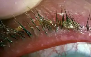
Contents [ hide ] Blepharitis is an inflammation of the eyelids often close to the base of the lashes. It usually occurs when tiny glands between lashes become blocked .  Blepharitis is the inflammation of the eyelids. The eyelid is folds of skin that keep the eye safe from debris and injury on the edge of the lids are lashes with short curved hair follicles containing oil glands. Blepharitis commonly occurs when the tiny oil glands located on the edge of the eyelids become clogged or irritated resulting in red eyes the condition usually occurs in both eyes and involves the part of the eyelid.  Where the eyelashes grow,  it is a common condition that can develop at any age but is more prevalent in young children and people older than 50 years of age. These blocks often produce a crusty or oily appearance with flaking of the skin around the eyelid. The lid themselves may become red and swollen. Blepharitis may cause a gritty burning or itchy sensation while uncom...

Chalazion(Etiology,symptoms,signs,complication,treatment)

Image
The tarsal or meibomian cyst is a chronic non-infective (non-suppurative) lipogranulomatous inflammation of the meibomian gland is called a chalazion . This is the commonest of all lid lumps. Glands of eyelids Meibomian glands Glands of Zeis Glands of moll Accessory lacrimal glands of wolverine Etiology Predisposing factors are similar to a hordeolum. Pathogenesis. First, there occurs mild grade infection of the meibomian gland by organisms of very virulence. The pent-up and extravasated secretion act like an irritant and excite non-infective  lipogranulomatous inflammation of the meibomian gland and surrounding tissue. Symptoms Painless swelling in the eyelid, gradually increases in size is the main presenting symptom. The Mild heaviness in the lid may be felt with moderately large chalazion. The Blurred vision may occur occasionally due to induced astigmatism by a very large chalazion pressing on the cornea. The Watering may occur sometimes due to eversion of the lower punctum ca...

Corneal scarring | Causes | Symptoms | Types | Treatment

Image
Corneal scarring This is the usual end result of healed corneal ulcer . The Corneal scarring leads to permanent visual impairment ranging from slight blurring to total blindness. Depending upon the clinical course of the ulcer, corneal scar noted maybe a nebula, macula, leucoma, ectatic cicatrix or keratectasia, adherent leucoma, or anterior staphyloma. It plays a key role in vision. The light that enters to eye gets refracted or bent by the cornea's curved edges. This help to determine how well your eye can focus on object close-up and far away. If the cornea is damaged by disease, injury, infection, the resulting scars will affect vision. They can block or distort light as it enters the eye.  It's actually one of the most common eye injuries and even though it can be painful, it's usually quite minor. It can happen for all kinds of reasons, from sports or DIY injuries to getting sand in your eye. Causes Anything that makes contact with the surface of the eye can scratc...

Hyperacute Bacterial Conjunctivitis | Etiology, Symptoms,Signs, Treatments

Image
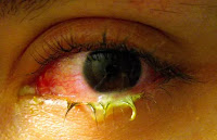
Hyperacute bacterial conjunctivitis is also known as acute purulent conjunctivitis or acute amenorrhea is characterized by a violent inflammatory response. It occurs in two forms Adult purulent conjunctivitis Ophthalmia neonatorum in newborn  Hyperacute Conjunctivitis of Adults  (Gonococcal conjunctivitis) Aetiology The disease affects adults, predominantly males, gonococcal infection directly spreads from genitals to eye. Presently, the incidence of gonococcal conjunctivitis has markedly decreased. Symptoms Pain which is moderate to severe. Purulent discharge, which is usually copious,  Swelling of eyelids, which is usually marked. Discomfort and foreign body sanitation Photophobia Sign Eyelids are tense and swollen  Tenderness is marked Discharge is thick purulent, copious trickling down the cheeks. Conjunctiva shows marked chemosis, congestion and papillae, giving bright red velvety appearance. Frequently, a pseudomembrane may be seen on the conjunctival surface. ...